Articles
Counterfeit detection of pharmaceutical tablets with transmission Raman spectroscopy
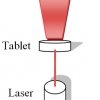
Counterfeiting is a major problem throughout the world and transmission Raman spectroscopy proves to be a very useful tool for fast and non-destructive detection of counterfeit pharmaceuticals.
Development of fibre-optic Raman probes for in vivo diagnosis of upper gastrointestinal cancers
This article surveys developments in the applications of Raman spectroscopy and the design of probes for use in endoscopes for the detection of early cancers in the throat and stomach.

The search for signatures of early life on Mars: Raman spectroscopy and the Exomars mission
Raman spectroscopy is to be used as one of the analytical techniques for the European Space Agency’s ExoMars mission to identify the geological and biogeological spectral signatures that could herald the presence of extinct or extant life on Mars. The article looks at the benefits of Raman spectroscopy for this and the research on Earth to build knowledge of the spectra of organisms living in extreme conditions here.
Towards clinical in vivo applications of Raman spectroscopy
Here, we focus on new trends in Raman spectroscopy to improve in vivo diagnosis. The use of Raman spectroscopy for real-time diagnosis of medical disease without the need for biopsy is among the most exciting and clinically relevant applications; four recent reports are presented. First, an approach to reduce fluorescent background of lung tissue in combination with a biomedical filtered Raman fibre optic probe was introduced in 2009 by Magee et al. Second, a fibre optic probe was developed for the CARS variant of Raman spectroscopy. Third, functional metal nanoparticles and carbon nanotubes were applied to a small animal model to collect Raman spectra non-invasively utilising the surface enhanced Raman scattering (SERS) effect. Finally, spatially offset Raman spectroscopy (SORS) has been presented as another non-invasive Raman-based method to probe deep bone subcutaneously in an animal model.
Simultaneous Raman-LIBS for the standoff analysis of explosive materials
The problem of detecting, recognising and identifying explosives at significant standoff distances has proved one of the most difficult—and most important—challenges during recent years, being today, one of the most demanding applications of spectroscopic techniques. The limited number of sophisticated available techniques potentially capable of standoff detection of minimal amounts of explosives is based on laser spectroscopy. Of the recently developed techniques, Raman spectroscopy and laser-induced breakdown spectroscopy (LIBS) are considered significant for their potential for homeland defence applications.

Complementary spectroscopic analyses of varnishes of historical musical instruments
For the past two centuries, the nature of the varnishes coating historical instruments has been a much debated subject. Focusing in particular on the varnishes used for coating violins made by the Italian instrument-maker Antonio Stradivari, numerous hypotheses have been raised by instrument-makers, experts, musicians and chemists, without reaching a general understanding of the ancient varnishing techniques. A few years ago, we decided to work on this topic using several complementary approaches for materials characterisation and study of historical sources (ancient varnish recipes, etc.).
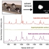
Universal Raman enhancement by solvent removal
Interest in Raman spectroscopy as an analytical technique that can be applied in a wide variety of fields continues to increase. The main reason for this interest is that no special sample preparation is required. However, the Raman signal is typically very weak, with only one in every 106–108 photons being scattered. This has driven the development of several enhancement techniques, e.g. Resonance Raman (RR), Surface Enhanced Raman Spectroscopy (SERS) and Surface Enhanced Resonance Raman Spectroscopy (SERRS), which can be used for dilute samples.
Detection of counterfeit erectile dysfunction drugs with Raman spectroscopy
This article will demonstrate the use of Raman spectroscopy as a fast and easy detection system for different counterfeit erectile dysfunction drugs. The possible counterfeits analysed consisted of two Viagra® tablets, SEYAGRA-GEL containing the same active ingredient as Viagra® and two tablets claiming to contain the same active ingredient as Cialis®.
Hidden depths? New techniques for sub-surface spectroscopy
A number of analytical applications in the area of security screening, medical diagnosis, drug authentication and quality control often require non-invasive probing of diffusely scattering (turbid) media in order to obtain chemical characterisation of deep-lying sample regions. Examples include non-invasive disease diagnosis, the detection of concealed explosives and illicit materials, the identification of counterfeit drugs and quality control applications in the pharmaceutical industry. Raman spectroscopy holds particular promise in this area due to its inherently high chemical specificity [exceeding that of near infrared (NIR) absorption spectroscopy and comparable with mid-infrared and THz methods], the ability to probe samples in the presence of water (the Raman scattering cross-section of water is very low) and its high penetration depth into turbid non-absorbing or weakly absorbing samples. On the downside, the technique is restricted to samples that do not exhibit strong fluorescence emission although this problem can, in the majority of cases, be avoided by using NIR excitation. Until recently, Raman techniques have generally been confined to applications involving surface layers of turbid media due to limitations imposed by the backscattering collection geometry common to the majority of commercial Raman probes. In principle, confocal Raman microscopy can potentially resolve objects to depths of up to several hundred micrometres. Deeper layers cannot be readily resolved and, typically, are overwhelmed by Raman and fluorescence signals emanating from the surface layer.

Raman spectroscopy in the forensic conservation of a unique marine artefact: the HMS Victory Trafalgar sail
Raman spectroscopy was used to study the condition of the Victory sail. Molecular spectroscopic analysis of the sail fibres was needed to simulate the aged, degraded material, thereby effecting a better compatibility between the old and replacement materials which would assist in the preservation of this ancient, historic marine textile.
Raman spectroscopy goes to Mars
Fernando Rull Pérez and Jesus Martinez-Frias
Centro de Astrobiología, Unidad Asociada CSIC-Universidad de Valladolid, Facultad de Ciencias, 47006 Valladolid, Spain
Raman spectroscopy: a simple, non-destructive way to characterise diamond and diamond-like materials
Jacob Filik
School of Chemistry, University of Bristol, Bristol, BS8 1TS, UK
Micro-spectroscopy: shedding light on rock formation
Simon FitzGerald
Horiba Jobin Yvon Ltd, 2 Dalston Gardens, Stanmore, Middlesex HA7 1BQ, UK. E-mail: [email protected]
Total internal reflection Raman spectroscopy
Phillip R. Greene and Colin D. Bain
Department of Chemistry, University of Oxford, Chemistry Research Laboratory, Mansfield Road, Oxford, OX1 3TA, UK
Raman analyses and "smart" imaging of nanophases and nanosized materials
Philippe Colomban
LADIR-UMR 7075 CNRS & Université P. & M. Curie, 2 rue Henry Dunant, 94320 Thiais, France
Infrared and Raman spectroscopic imaging in biosciences
David Chenery and Hannah Bowring
Smith & Nephew Group Research Centre, York Science Park, Heslington, York YO10 5DF, UK
Raman spectroscopy: about chips and stress
Moore’s law* dictates microelectronics researchers to make integrated circuit (IC) devices smaller and to put them as close to each other as possible on a chip. This results in a better performance and a larger functionality of the chips. However, these devices also require a good electrical isolation from each other. This is in general done by the formation of a thick local oxide in the “field region” between the devices. In 1970, researchers from Philips1 invented the so-called LOCOS (LOCal Oxidation of Silicon) technique to achieve this isolation. Using a Si3N4 mask, the silicon is thermally oxidised in the nitride-free field regions. Figure 1 (left) shows a typical LOCOS structure. Although LOCOS seemed a perfect solution at that time, it came with a lot of problems, many of them related to mechanical stresses. Thermal oxidation of Si to SiO2 occurs together with a 125% volume expansion. As a result, the oxide grown in the field region, called the “field oxide,” exerts large forces on the surrounding silicon. Another major drawback of this technique is the so-called “bird’s beak,” caused by the lateral growth of the oxide under the nitride mask. This bird’s beak not only affects the intended device length, it also introduces large local mechanical stresses in the silicon, because of volume expansion, and it also deforms the nitride film. These stresses often resulted in the generation of dislocations in the silicon, which are quite harmful for the devices.
Extracting Raman spectra from highly fluorescent samples with "Scissors" (SSRS, Shifted-Subtracted Raman Spectroscopy)
S.E.J. Bell,a* E.S.O. Bourguignon,a A. O’Grady,a J. Villaumiea and A.C. Dennisb
aSchool of Chemistry, The Queen’s University of Belfast, Belfast, BT9 5AG, Northern Ireland, UK
bAvalon Instruments Ltd, 10 Malone Road, Belfast BT9 5BN, Northern Ireland, UK
Micro-Raman technique for phase analysis on archaeological ceramics
The study of the mineralogical phases of archaeological ceramics may be very helpful in unravelling the history of an ancient sherd, particularly by means that investigate the process of its production. Micro-Raman spectroscopy offers advantages as a non-destructive, or even better, a non-sampling technique.