Articles
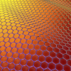
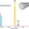
Graphene characterisation and standardisation via Raman spectroscopy
Graphene has been receiving a large amount of interest as its commercial possibilities begin to be realised. Now, with hundreds of companies offering commercial graphene production, analytical measures of graphene quality are required. Raman spectroscopy can be used to “understand the number of layers, strain, doping and importantly the level of disorder present in graphene”, which is described in this article: “Graphene characterisation and standardisation via Raman spectroscopy” by Andrew Pollard and Debdulal Roy.
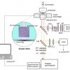
Automated detection of counterfeit drugs using multimodal spectroscopy and advanced web-based software platforms
In this Tony Davies Column, we learn about “Automated detection of counterfeit drugs using multimodal spectroscopy and advanced web-based software platforms”. The German authorities have commissioned the development of a multi-modal, transportable inspection system, including intelligent data processing and evaluation, for fast spectroscopic recognition of illicit drugs and counterfeit medicines. This is described in the column.
Monitoring of catalytic reactions and catalyst preparation processes in liquid phase systems by combined in situ spectroscopic methods
In situ spectroscopic methods such as infrared, Raman and UV/vis spectroscopy are powerful tools to gain insight into reaction mechanisms and catalyst actions in homogeneously catalysed reactions. These methods and combinations of them offer great potential for the real-time monitoring of reactions in the liquid phase, for mechanistic studies as well as process control and kinetics.
In vivo Raman spectroscopy of skin
“In vivo Raman spectroscopy of skin” is Paul Pudney’s topic. The skin is a most important part of our bodies. There is great interest in studying it to help understand the many skin diseases we are prone to, including cancer, to develop skin care products and, increasingly, as an alternative route to administer pharmaceuticals instead of through the gut. Raman spectroscopy is an excellent tool to study these, and has particular advantages in its ability to do so in vivo.
Application of Raman and photoluminescence spectroscopy for identification of uranium minerals in the environment
The “Application of Raman and photoluminescence spectroscopy for identification of uranium minerals in the environment” is described by Eric Faulques, Florian Massuyeau, Nataliya Kalashnyk and Dale Perry. Uranium forms a large number of compounds and complexes, and these are most helpful in the study of uranium, its chemistry and transport in the environment. Raman and photoluminescence spectroscopy provide complementary information and are powerful tools for direct speciation of uranium and identification of natural uranyl minerals relevant to the environment.

Optical spectroscopy in therapy response monitoring: an awakening giant
“Optical spectroscopy in therapy response monitoring: an awakening giant” by Arja Kullaa, Surya Singh, Jopi Mikkonen and Arto Koistinen looks at the important advances made by optical spectroscopy techniques, such as diffuse optical spectroscopic imaging (DOSI), Raman, diffuse reflectance and fluorescence spectroscopy, in changing how cancer is managed in patients. The ability to repeatedly monitor tumour dynamics to see how effective a particular treatment has been has enormous potential for us all.
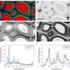
Elucidating structural and compositional changes in plant tissues and single cells by Raman spectroscopic imaging
“Elucidating structural and compositional changes in plant tissues and single cells by Raman spectroscopic imaging” is the topic of the next article by Batirtze Prats Mateu, Barbara Stefke, Marie-Theres Hauser and Notburga Gierlinger. Understanding plant cells is important for the best use of plants in traditional and new applications. Raman spectroscopic imaging represents one of the best ways to unravel the molecular structure in the native environment of plant tissues.

Emerging sampling approaches for Raman analysis of foods
“Emerging sampling approaches for Raman analysis of foods” by Nils Kristian Afseth, Matthew Bloomfield, Jens Petter Wold and Pavel Matousek describes how a number of instrumental developments are enabling Raman spectroscopy to find increasing applications in food analysis. They show how Spatially Offset Raman Spectroscopy (SORS) is being used to analyse quality parameters in salmon, including the content of fat, its fat composition and the content of carotenoids. Traditionally, the preserve of NIR spectroscopy, Raman may increasingly be used for the analysis of food and other biological matrices.
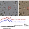
Raman spectroscopy for the study of biological organisms (biogenic materials and biological tissues): a valuable analytical tool
Malvina G. Orkoulaa and Christos G. Kontoyannisa,b
aDepartment of Pharmacy, University of Patras, Rio-Patras, Greece. E-mail: [email protected]
bICE-HT/FORTH, PO Box 1414, University Campus, Rio-Patras, Greece
Probing and sorting single cells: the application of a Raman-activated cell sorter
The single cell Raman spectrum (SCRS) enables cell probing and sorting to study phenotypes and ecophysiology of single cells and explore individual cells in situ in a label-free and non-destructive manner.
Highlighting the importance of utilising the polarisation properties of resonance Raman scattering in obtaining molecular information
In resonance Raman scattering (RRS), the amount of structural and chemical information deduced can be increased by analysing the polarisation of the inelastically scattered light, including the degree of molecular aggregation in bio-molecules in their natural environment.
Detection of thiabendazole applied to organic fruit by near infrared surface-enhanced Raman spectroscopy
Thiabendazole (TBZ) is a chemical fungicide and parasiticide used to prevent mould, blight and other diseases resulting from long transportation and storage, largely used as an ingredient in waxes applied to the skins of citrus fruits. The authors describe their work using near infrared-surface-enhanced Raman spectroscopy and conventional Ag nanoparticles, which showed that TBZ was found both on organic fruit and at levels higher than regulations allow.
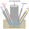
Tip-enhanced Raman mapping (TERM) of single-walled carbon nanotubes and graphene
Tip-enhanced Raman spectroscopy (TERS) and tip-enhanced Raman mapping (TERM) can be used advantageously to investigate the carbon allotropes graphene and single-walled carbon nanotubes, with a spatial resolution in the nanometre range
Surface-enhanced Raman scattering (SERS) spectroscopy identifies fraudulent uses of fuels
Fuels and the taxes raised from their sale are a big business around the world. To control smuggling, counterfeiting, theft and product diversion, markers can be placed in the fuel. The use of a SERS active compounds as markers is described as well as the development of a portable instrument for detection of the markers in the field.
Representative Raman measurements of carbon nanotubes
This article explains what is represented in a Raman spectrum of carbon nanotubes and how to optimise the measurement. There is actually significant diversity within samples of nanotubes which affects both the material properties and the Raman spectrum of the material.

An important use of Raman spectroscopy to help understand the impact of traffic on roadside soils and plants
Even though lead in fuel has been banned for a number of years, it is still present in by the roadside, as are many other pollutants from vehicles. The combination of Raman spectroscopy and µ-ED-XRF is of particular value. The advantage Raman has is in the possibility of focusing on individual grains, thereby obtaining the spectrum of each grain that comes from traffic-emitted particles.
Following lipids in the food chain: determination of the iodine value using Raman micro-spectroscopy
Raman spectroscopy is used to monitor the iodine value of algal-derived fish feed for its lipid content and to monitor algae samples for biofuel production.
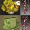
Fireworks: composition and chemistry through Raman spectroscopy and SEM-EDS imaging
Whilst fireworks are a great entertainment, they can also be used for illegal activities as well as potentially containing dangerous chemicals. The combination of Raman spectroscopy and SEM-EDS turns out to be a very efficient analytical method. In fact, these complementary techniques may also be used to analyse other kinds of pyrotechnic artefacts, low explosive formulations, high explosives, explosion residues etc.
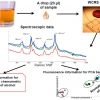
On-chip chemical fingerprinting of an analyte using both Raman spectroscopy and fluorescence
Praveen Ashok and Kishan Dholakia of St Andrews University, UK, describe the scope of optofluidic devices that can be implemented using the waveguide confined Raman spectroscopy (WCRS) technique they have developed. I am particularly impressed by the sample size of whisky shown in Figure 2—true Scottish style!
Time-resolved Raman spectroscopy for non-invasive detection through non-transparent materials
Since attempts to blow up planes using liquid explosives, we have all been restricted in what we can take onboard when we fly. Raman spectroscopy is offering a solution. However, “Time-resolved Raman spectroscopy for non-invasive detection through non-transparent materials” by Ingeborg Iping Petterson and Freek Ariese argues that time-resolved Raman spectroscopy (TRRS) techniques provide better spatial selectivity than the major alternative, spatially offset Raman spectroscopy (SORS). Applications for through-skin measurements and depth analysis in catalytic extrudates are also described.