Articles
Multisensor hyperspectral imaging as a versatile tool for image-based chemical structure determination
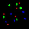
The authors describe “Multisensor hyperspectral imaging as a versatile tool for image-based chemical structure determination”. They describe the features of a software package that allows the combined analysis of hyperspectral data from different imaging techniques. This multisensor approach providing complementary information has many advantages.
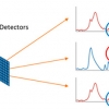
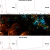
Quantum cascade laser-based mid-infrared spectrochemical imaging of tissues and biofluids
Mid-infrared spectroscopic imaging is a rapidly emerging technique in biomedical research and clinical diagnostics that takes advantage of the unique molecular fingerprint of cells, tissue and biofluids to provide a rich biochemical image without the need for staining. Spectroscopic analysis allows for the objective classification of biological material at a molecular level.1 This “label free” molecular imaging technique has been applied to histology, cytology, surgical pathology, microbiology and stem cell research, and can be used to detect subtle changes to the genome, proteome and metabolome.2–4

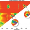
Atomic dielectric resonance
It is important that places of archaeological and architectural importance need to be explored without damage. Atomic Dielectric Resonance (ADR) can be used for identifying sub-surface geological features. This technology1 uses a novel coherent beam, which has been used in the oil, gas and water industries, to provide information on what lies beneath the earth’s surface, without the need to drill cores.
Review of nanoscale infrared spectroscopy applications to energy related materials
The authors give us a “Review of nanoscale infrared spectroscopy applications to energy related materials”. Fuel cells, photovoltaics and specialised polymers for fracking are all considered.
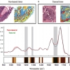
Novel concepts in infrared spectral imaging as a cancer diagnostic tool
This article describes an application of spectral imaging for the differentiation of tumour and normal cells. The authors also introduce the concept of a spectral barcode, which has had success with some tissues and has potential in others.
Near infrared hyperspectral imaging for foreign body detection and identification in food processing
With continuing food scares around the world, food producers need every tool they can get to prevent contamination of their products at every stage of production. Hyperspectral reflectance imaging in the NIR combined with chemometrics shows much promise for the detection and identification of foreign bodies among food grains.
Fourier transform infrared spectroscopic imaging of live cells
There are a number of approaches, and by combining FT-IR imaging methodology with microfluidics devices, the opportunity to study live cells by FT-IR imaging in controlled environments is now possible.
Single particle characterisation in biologics: from mid-infrared micro-spectroscopy and mapping to spectral imaging
The presence of “particles” in protein pharmaceuticals (biologics) can cause severe, unwanted effects in the drug. The article describes the use of mid-infrared micro-spectroscopy for the investigation and chemical characterisation of single particles in these biologics.
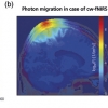
Measuring brain activity using functional near infrared spectroscopy: a short review
“Measuring brain activity using functional near infrared spectroscopy: a short review” by Felix Scholkmann and Martin Wolf looks at the various methods for performing fNIRS and some applications that demonstrate why this non-invasive, safely applicable, portable and cost-effective method is now an integral part of the techniques used in neuroscience.
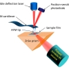
Reverse engineering of polymeric multilayers using AFM-based nanoscale IR spectroscopy and thermal analysis
It is possible to obtain both infrared spectra and thermal analysis data of individual layers in a cross-sectioned multilayer film. Since both techniques are AFM-based, the topographical features can be readily linked to the spectroscopic and thermal data at a much higher spatial resolution than previously achievable.
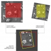
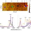
Fireworks: composition and chemistry through Raman spectroscopy and SEM-EDS imaging
Whilst fireworks are a great entertainment, they can also be used for illegal activities as well as potentially containing dangerous chemicals. The combination of Raman spectroscopy and SEM-EDS turns out to be a very efficient analytical method. In fact, these complementary techniques may also be used to analyse other kinds of pyrotechnic artefacts, low explosive formulations, high explosives, explosion residues etc.

The role of microspectroscopy techniques in the study of historic artworks
Nati Salvadó, Salvador Butí and Trinitat Pradell have used a number of techniques to investigate changes in pictorial techniques in Catalan paintings in the 15th century. The combination of different techniques is of particular value. The use of synchtrotron radiation as a light source is also an advantage.
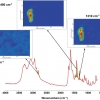
Revealing the presence of creatine in human spinal cord in amyotrophic lateral sclerosis, by infrared microspectroscopy
We have previously investigated the topographic and quantitative changes in the distribution of trace metals in spinal cords from ALS and control patients. X-ray fluorescence microscopy was used to investigate their metallic nature and distribution in single nerve cells. A deeper understanding of the neurodegenerative processes in ALS requires focus on the biochemical changes occurring in nervous tissue of such a disorder. For this purpose, we have undertaken an infrared microspectroscopy study. While metals are suggested to play a pivotal role in the pathogenesis of ALS, they typically do not occur in tissues as free ions. This results in the presence of the complex mechanisms of metal ions buffering that protect cells against their toxic effects. Metal homeostasis is regulated by several proteins. Such proteins containing metal cofactor are called metalloproteins.
Laser ablation ICP atomic emission spectrometry: a new tool for imaging of pharmaceutical tablets
Imaging of organic and inorganic constituents of tablets represents a considerable challenge and no single spectroscopic approach can provide definitive characterisation of all components and/or satisfy key measurement criteria such as sensitivity, specificity, resolution and speed of analysis. Laser ablation in combination with ICP emission spectrometry represents a powerful new tool for imaging elemental distribution in pharmaceutical tablets.
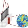
The sound of science
I recently “discovered” a very interesting radio programme on BBC Radio 4. It is “devoted to the powerful, sometimes beautiful, often abused but ever ubiquitous world of numbers”. A few weeks ago we were asked to say what we were doing while listening to the programme. The next week we were told that nearly 2000 e-mails had been received and this data had been given to information designer David McCandless to turn into a graphic. When this was trailed I got the impression that something new and exciting was going to be displayed and I thought that the graphic would include sound. The graphic is good but rather “ordinary” and I was disappointed. This got me thinking about how we display information. Have we made any advance in the last 25 years? Could sound be used!
The role of MALDI-enabled linear ion trap mass spectrometry as a sensitive tool in tissue imaging
This article discusses matrix-assisted laser desorption/ionisation (MALDI) enabled linear ion trap (LIT) mass spectrometry (MS) as a technique for fast and accurate tissue imaging, compared to the more traditional time-of-flight (ToF) method.
XPS surface analysis: imaging and spectroscopy of metal and polymer surfaces
Antonella Rossi,a,b Bernhard Elsenera and Nicholas D. Spencerb aDepartment of Inorganic and Analytical Chemistry, University of Cagliari, Campus of Monserrato, 09100 Cagliari, Italy
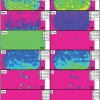
Polymer characterisation by spectroscopic imaging: infrared and electron microscopy: a good match
Peter Wilhelm,a Boril Chernev,a Peter Pölt,a Gerald Kothleitner,a Klaus-Jochen Eichhorn,b Gisela Pompe,b Nikola Johnerc and Alexander Piryc
aResearch Institute for Electron Microscopy, Graz University of Technology; Steyrergasse 17, A-8010 Graz, Austria. E-mail: [email protected]
bInstitute of Polymer Research Dresden; Hohe Straße 6, D-01069 Dresden, Germany
Near infrared spectroscopy: the practical chemical imaging solution
Chemical imaging spectroscopy is an exciting new analytical advance that answers commonly asked questions such as what chemical species are in a sample, how much of each is present, and most importantly, where are they located? Through the fusion of traditional infrared spectroscopy with powerful microscopic and macroscopic imaging capabilities, chemical imaging spectroscopy answers all these questions simultaneously, in a single rapid measurement.