Jürgen Gross has been using ambient mass spectrometry to look at the presence of polydimethylsiloxanes (PDMS) in food prepared in silicone rubber objects and on baking parchment. He shows that PDMS migrates into the food, something perhaps we should think about if in the mood for some baking!
Spectroscopy Articles
Displaying 81–100 of 302 results
Pages

Issue 27/4 (2015)

Stanislav Strekopytov tells us about “The use of inductively coupled plasma mass spectrometry to quantify chemical hazards in natural history collections: arsenic and mercury in taxidermy bird specimens”. It is quite shocking to learn about the use of poisons to preserve taxidermy specimens in the past. Nowadays it is essential that the dangers from such specimens are known before they can be handled by museum staff and particularly if they might be touched by visitors. ICP-MS analysis provides fully quantitative information on bulk contents of toxic elements in taxidermy specimens and so is well suited to this task.
Issue 27/4 (2015)

Mark Tobin and colleagues describe “Fourier transform infrared spectroscopy and imaging of dragonfly, damselfly and cicada wing membranes”. Insects and plants have evolved highly specialised surfaces such as being highly water repellent or superhydrophobic, which also confers self cleaning properties. This is of interest to materials scientists to help in the development of manufactured materials with similar properties. High spatial resolution FT-IR spectroscopy and imaging provide useful information about the complex chemical patterning that contributes to this functionality.
Issue 27/4 (2015)
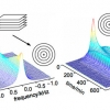
The authors describe “Infrared mapping spectroscopic ellipsometry”. Recent developments in fundamental and materials research have increased the value of mapping techniques such as ellipsometry. IR ellipsometry, since it operates in the mid-IR fingerprint region, provides complementary information on composition, structural properties and interactions
Issue 27/3 (2015)
In situ spectroscopic methods such as infrared, Raman and UV/vis spectroscopy are powerful tools to gain insight into reaction mechanisms and catalyst actions in homogeneously catalysed reactions. These methods and combinations of them offer great potential for the real-time monitoring of reactions in the liquid phase, for mechanistic studies as well as process control and kinetics.
Issue 27/3 (2015)

Research is under way as to the possibility of using high-precision isotopic analysis of metals in a biomedical context. The goal is to develop methods for medical diagnosis on the basis of isotopic analysis of mineral elements in biofluids, for diseases that can otherwise only be established at a later stage or via a more invasive method (e.g., a biopsy) and/or for prognosis purposes. Whilst this work is in a very early stage, it is known that various diseases have an influence on the uptake, metabolism and/or excretion of essential mineral elements and thus, can cause a difference in their isotopic composition in biofluids.
Issue 27/3 (2015)

“In vivo Raman spectroscopy of skin” is Paul Pudney’s topic. The skin is a most important part of our bodies. There is great interest in studying it to help understand the many skin diseases we are prone to, including cancer, to develop skin care products and, increasingly, as an alternative route to administer pharmaceuticals instead of through the gut. Raman spectroscopy is an excellent tool to study these, and has particular advantages in its ability to do so in vivo.
Issue 27/2 (2015)

Christian Schröder tells us about “Mössbauer spectroscopy in astrobiology”. Iron is abundant in the Earth’s crust, as well as on Mars and is likely to be so also on Jupiter’s moon, Europa. Iron is important for life and may have played a role in the origin of life as an energy source and by providing mineral surfaces as a template for surface metabolism. Iron continues to be essential for almost all organisms as the functional centre of many proteins and enzymes. Mössbauer spectroscopy is a powerful tool to study iron-bearing solid substances and as such has applications in the search for life in other parts of our Solar System.
Issue 27/2 (2015)
Sampling on important works of art is not possible and this is the main reason why only non-invasive techniques, such as MSI, are becoming increasingly popular to assist with undertaking conservation decisions.
Issue 27/2 (2015)

Yvonne Fors, Håkan Grudd, Anders Rindby and Lennart Bornmalm tell us about “X-ray fluorescence for cultural heritage: scanning biochemical fingerprints in archaeological shipwrecks”. Two outstanding examples of the preservation of wood are the warships Vasa, in Stockholm and the Mary Rose in Portsmouth and this article looks at the role XRF has played in the preservation of the wood of both ships.
Issue 27/1 (2015)
The “Application of Raman and photoluminescence spectroscopy for identification of uranium minerals in the environment” is described by Eric Faulques, Florian Massuyeau, Nataliya Kalashnyk and Dale Perry. Uranium forms a large number of compounds and complexes, and these are most helpful in the study of uranium, its chemistry and transport in the environment. Raman and photoluminescence spectroscopy provide complementary information and are powerful tools for direct speciation of uranium and identification of natural uranyl minerals relevant to the environment.
Issue 27/1 (2015)

“Optical spectroscopy in therapy response monitoring: an awakening giant” by Arja Kullaa, Surya Singh, Jopi Mikkonen and Arto Koistinen looks at the important advances made by optical spectroscopy techniques, such as diffuse optical spectroscopic imaging (DOSI), Raman, diffuse reflectance and fluorescence spectroscopy, in changing how cancer is managed in patients. The ability to repeatedly monitor tumour dynamics to see how effective a particular treatment has been has enormous potential for us all.
Issue 27/1 (2015)
The authors tell us about “Two dimensional elemental mapping by laser-induced breakdown spectroscopy”. LIBS seems to be finding increasing applications and to be receiving interest by the instrument manufacturers at present. The article provides an introduction to the technique and goes on to show how it can be used for elemental mapping in materials analysis.
Issue 26/6 (2014)
“Rheo-nuclear magnetic resonance spectroscopy: a versatile toolbox to investigate rheological phenomena in complex fluids” is Claudia Schmidt’s topic. Rheology is an important science, and NMR has a number of uses within it. However, challenges remain for the simultaneous measurement of rheological and NMR parameters.
Issue 26/6 (2014)
“Elucidating structural and compositional changes in plant tissues and single cells by Raman spectroscopic imaging” is the topic of the next article by Batirtze Prats Mateu, Barbara Stefke, Marie-Theres Hauser and Notburga Gierlinger. Understanding plant cells is important for the best use of plants in traditional and new applications. Raman spectroscopic imaging represents one of the best ways to unravel the molecular structure in the native environment of plant tissues.
Issue 26/5 (2014)
The authors describe “Multisensor hyperspectral imaging as a versatile tool for image-based chemical structure determination”. They describe the features of a software package that allows the combined analysis of hyperspectral data from different imaging techniques. This multisensor approach providing complementary information has many advantages.
Issue 26/5 (2014)
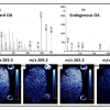
Robert Bradshaw and Simona Francese tell us about “Matrix–assisted laser desorption ionisation tandem mass spectrometry imaging of small molecules from latent fingermarks“. Especially when looking at small molecules in fingermarks, isobaric species can be a problem and this has the potential to affect the outcome of any court case if not handled appropriately. Tandem mass spectrometry can be used as an alternative to high-resolution MS and ion mobility.
Issue 26/4 (2014)
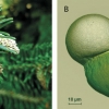
Currently, pollen identification is mostly done under a light microscope. FT-IR spectroscopy of pollen grains provides rapid and simple identification of pollen, with the added benefit of providing environmental information.
Issue 26/4 (2014)
Orthogonal spectroscopic techniques for the early developability assessment of therapeutic protein candidates” are described by Patrick Garidel, Anne Karow and Michaela Blech. Due to its cost and time implications, in the early development phase of drug discovery the use of othogonal techniques, based on different physical observables, is important for correct decision-making.
Issue 26/4 (2016)
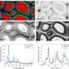
Mid-infrared spectroscopic imaging is a rapidly emerging technique in biomedical research and clinical diagnostics that takes advantage of the unique molecular fingerprint of cells, tissue and biofluids to provide a rich biochemical image without the need for staining. Spectroscopic analysis allows for the objective classification of biological material at a molecular level.1 This “label free” molecular imaging technique has been applied to histology, cytology, surgical pathology, microbiology and stem cell research, and can be used to detect subtle changes to the genome, proteome and metabolome.2–4
Issue 26/4 (2014)